Disc edge veins of Kraupa associated with optic disc drusen
Andrea Díaz 1,2Miguel Angel Almela 1
1 Department of Ophthalmology, Hospital Lluís Alcanyís, Xàtiva (Valencia), Spain
2 E.B.O. European Boarding of Ophthalmology
Abstract
Objective: Disc edge veins of Kraupa are a rare anomaly of the retinal venous system in which the main trunk of the retinal vein disappeared into the margin of the optic disc instead of its centre.
Methods: A 40-year-old woman was detected to have an anomaly in her left optic disc in a routine eye examination. The eyes had an anomaly of the retinal venous system in which all branches of the retinal vein joined in a common trunk that entered the disc margin inferonasally. The central retinal artery issued from the centre of the disc separately of the venous system. B-scan ultrasonografhy revealed the presence of hyperechoic imaging at the optic nerve head in both eyes.
Results: We describe the association of disc edge veins of Kraupa with optic disc drusen.
Conclusion: Vascular complications of optic disc drusen hav been described. We don’t know the implication of disc edge veins in the pathogenesis of these complications.
Introduction
In 1915, Ernst Kraupa published a drawing and description of a rare anomaly of the retinal venous system in which the main trunk of the retinal vein disappeared into the margin of the optic disc instead of its centre. We report the fluorescein angiographic and fundus photographs findings in a patient with a rare exit anomaly of the retinal vein at the optic disc margin associated with optic disc drusen.
Case description
A 40-year-old woman came to our centre because she was detected to have an anomaly in her left optic disc in a routine eye examination. Our evaluation showed best-corrected vision of 20/20. The anterior segment was normal. Applanation pressures were 17 mmHg in both eyes. There was not a relative afferent pupillary defect.
The ocular fundi revealed that both discs were normal in diameter.
The right disc had an anomaly of the retinal venous system in which all branches of the retinal vein joined in a common trunk that entered the disc margin inferonasally at 4 o'clock (Figure 1 [Fig. 1], A). The central retinal artery issued from the centre of the disc separately of the venous system.
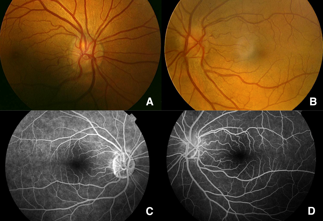
The left disc had an absence of cupping with blurred aspect of his margin. All retinal venous branches converged into a common venous trunk that entered the disc margin nasally at 11 o’clock (Figure 1 [Fig. 1], B). The central retinal artery issued from de centre of the disc temporally to the retinal vein.
Fluorescein angiography of the right eye (Figure 1 [Fig. 1], C) and the left eye (Figure 1 [Fig. 1], D), standard automated perimetry (Humphrey 24-2 test pattern on the Humphrey Visual Field Analyzer) and B-scan ultrasonography were performed.
Visual fields showed an enlargement of the blind spot in both eyes.
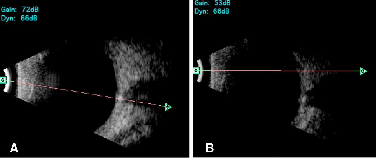
B-scan ultrasonografhy revealed the presence of hyperechoic imaging at the optic nerve head in the right eye (Figure 2 [Fig. 2], A) and in the left eye (Figure 2 [Fig. 2], B). These findings were considered consistent with disc edge veins of Kraupa associated with optic disc drusen.
Discussion
The ophthalmoscopic appearance of disc edge veins draining the retina has been previously reported [1]. We describe the association of this disc anomaly with optic disc drusen. In one hand, congenitally abnormal disc vasculature, such as abnormal branching pattern, presence of relatively large blood vessels connecting the superficial and deep disc circulations, and increased disc capillarity, may contribute to drusen formation. In the other hand, optic disc with drusen are often accompanied by irregular vascular pattern (ie, cilioretinal arteries and retinal choroidal venous collaterals) [2]. In our case, although the appearance of disc edge veins was bilaterally, the imaging of optic disc edema was only in the left eye, but the B-scan ultrasonography revealed the presence of buried drusen in both eyes.
Conclusion
Vascular complications of optic disc drusen such as optic disc and retinal hemorrhage, anterior ischemic optic neuropathy (AION), central retinal artery occlusion (CRAO), central retinal vein occlusion, and peripapillary choroidal neovascularisation [3], [4], [5] have been described.
We don’t know the implication of disc edge veins in the pathogenesis of these complications.
Notes
Patient’s consent
The patient has consented to the submission of the case report to the journal.
Competing interests
All authors certify that they have no affiliations with or involvement in any organization or entity with any financial interest (such as honoraria; educational grants; participation in speakers’ bureaus; membership, employment, consultancies, stock ownership, or other equity interest; and expert testimony or patent-licensing arrangements), or non-financial interest (such as personal or professional relationships, affiliations, knowledge or beliefs) in the subject matter or materials discussed in this manuscript.
References
[1] Barroso L, Hoyt WF, Narahara M. Disc edge veins of Kraupa: rare exit anomalies of the retinal vein. Br J Ophthalmol. 1992 Jul;76(7):442-3. DOI: 10.1136/bjo.76.7.442[2] Lam BL, Morais CG Jr, Pasol J. Drusen of the optic disc. Curr Neurol Neurosci Rep. 2008 Sep;8(5):404-8. DOI: 10.1007/s11910-008-0062-6
[3] Munteanu M. Complications hémorragiques des druses de la papille [Hemorrhagic complications of drusen of the optic disk]. J Fr Ophtalmol. 2007 Jan;30(1):58-67. DOI: 10.1016/S0181-5512(07)89552-0
[4] Sanders TE, Gay AJ, Newman M. Hemorrhagic complications of drusen of the optic disk. Am J Ophthalmol. 1971 Jan;71(1 Pt 2):204-17. DOI: 10.1016/0002-9394(71)90391-6
[5] Aumiller MS. Optic disc drusen: complications and management. Optometry. 2007 Jan;78(1):10-6. DOI: 10.1016/j.optm.2006.07.009

